Rare non-progressive retinal vasculopathy
reh-tuh-nuhl vas-kuh-lop-uh-thee
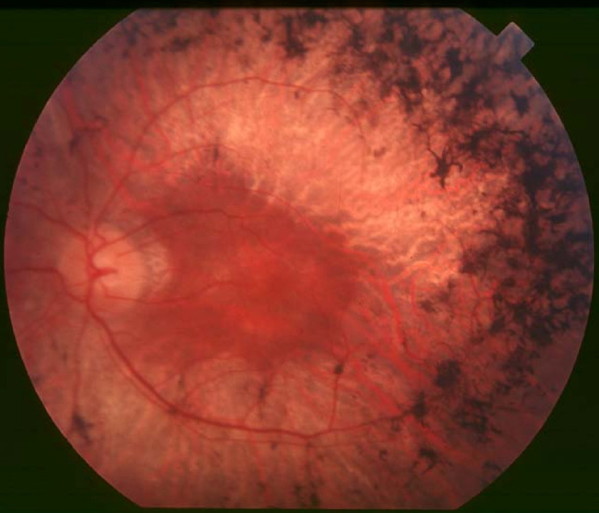
Also known as: retinal cavernous hemangioma, retinal racemose hemangioma
At a Glance
What is Rare non-progressive retinal vasculopathy?
Rare non-progressive retinal vasculopathy is a condition affecting the blood vessels in the retina, the light-sensitive tissue at the back of the eye. It is usually present at birth and does not worsen over time. The condition is caused by abnormal blood vessel formation in the retina. Early symptoms may include vision disturbances or floaters, while later symptoms can lead to more significant vision impairment. Early diagnosis is crucial to monitor and manage potential complications, such as retinal vein occlusion. The condition can impact family life by requiring regular eye examinations and potential interventions. Prognosis is generally good as the condition is non-progressive, meaning it does not worsen over time. Daily life for affected individuals may involve adapting to vision changes and attending regular ophthalmologic check-ups. Although it is a rare condition, awareness and monitoring can help manage symptoms effectively. The condition does not typically affect other body systems. Patients may need to be cautious about activities that could impact their vision. Support from healthcare professionals and family can help manage the condition effectively.
Medical Definition
Rare non-progressive retinal vasculopathy is characterized by the presence of abnormal retinal blood vessels, often referred to as cavernous hemangiomas. Histologically, these lesions consist of thin-walled, dilated vascular spaces filled with blood. The condition is classified under retinal vascular disorders and is considered non-progressive, meaning it does not typically lead to worsening over time. Epidemiologically, it is a rare condition with a prevalence of approximately 1 in 500,000 individuals. The disease course is generally stable, although complications such as retinal vein occlusion can occur. Management focuses on monitoring and addressing any complications that arise.
Rare non-progressive retinal vasculopathy Symptoms
Symptoms vary in severity between individuals. Early diagnosis and management can significantly improve outcomes.
Very Common
Visual disturbances manifest as blurred vision or visual field defects, often noticed during routine activities like reading or driving. These symptoms are caused by abnormal blood vessel formation in the retina, leading to altered blood flow and potential hemorrhages. Over time, these disturbances may stabilize or fluctuate, depending on the extent of vascular involvement. Daily life can be significantly affected, necessitating regular ophthalmologic evaluations and potential interventions to manage symptoms.
Floaters appear as small, moving spots in the field of vision, often described as cobwebs or specks. They result from small hemorrhages or debris within the vitreous humor due to retinal vascular changes. Floaters may increase in number or size over time, potentially indicating worsening of the underlying condition. Patients may find them distracting, especially against bright backgrounds, and should seek regular monitoring to assess changes.
Photopsia is experienced as flashes of light or flickering in the peripheral vision. This occurs due to mechanical stimulation of the retina by abnormal blood vessels or vitreous traction. The frequency and intensity of photopsia can vary, sometimes heralding more serious complications like retinal detachment. Patients are advised to report new or worsening episodes promptly to their eye care provider for assessment.
Common
Mild eye discomfort may present as a sensation of pressure or irritation in the affected eye. This discomfort is often due to the vascular changes and potential inflammation within the retina. While it may remain mild, it can occasionally escalate, particularly if complications arise. Patients can manage this symptom with over-the-counter pain relief and should maintain regular follow-ups to monitor any progression.
Decreased night vision is characterized by difficulty seeing in low-light conditions, such as driving at night. This symptom arises from impaired retinal function due to disrupted blood supply and potential retinal damage. Over time, night vision may deteriorate further, impacting the patient's ability to perform tasks in dim environments. Adaptive strategies, such as using brighter lighting and avoiding night driving, can help mitigate daily challenges.
Color vision changes involve difficulty distinguishing between certain colors or a general dulling of color perception. These changes are linked to retinal damage affecting the cones, the photoreceptors responsible for color vision. The progression of color vision changes can vary, potentially leading to more pronounced deficits. Patients may need to adapt by using color-coded aids and should have regular assessments to monitor changes.
Less Common
Peripheral vision loss is the reduction of side vision, often unnoticed until it affects daily activities. It occurs due to damage to the outer areas of the retina, where abnormal blood vessels may cause localized ischemia. This loss can progress slowly, leading to tunnel vision if untreated. Patients may require orientation and mobility training to adapt to changes in their visual field.
Headaches may occur as a secondary symptom, often due to eye strain or increased intraocular pressure. The underlying retinal changes can lead to discomfort and tension, contributing to headache development. These headaches can vary in intensity and frequency, sometimes correlating with visual disturbances. Management includes addressing the primary ocular issues and using standard headache treatments as needed.
What Causes Rare non-progressive retinal vasculopathy?
Rare non-progressive retinal vasculopathy is primarily associated with mutations in the KRIT1 gene located on chromosome 7q21. The KRIT1 gene encodes a protein that is crucial for maintaining the integrity of blood vessel walls. Mutations in KRIT1 lead to structural changes in the protein, impairing its ability to interact with other proteins involved in endothelial cell junctions. This disruption causes increased vascular permeability and abnormal blood vessel formation. As a result, there is a breakdown in the blood-retinal barrier, leading to localized retinal edema and hemorrhage. The accumulation of fluid and blood in the retinal layers can trigger a localized inflammatory response, involving microglial activation and cytokine release. This inflammation can exacerbate tissue damage and contribute to the degeneration of retinal structures. The pattern of symptoms, such as visual disturbances, is due to the specific retinal regions affected by the vascular anomalies. Variability in disease severity among patients can be attributed to differences in the extent of genetic mutations and their impact on protein function. Additionally, the presence of other genetic or environmental factors may influence the clinical presentation. In some cases, compensatory mechanisms within the retina may mitigate the effects of the mutations, leading to milder symptoms. The non-progressive nature of the condition suggests that once the initial vascular changes stabilize, further deterioration is limited. Understanding the precise molecular pathways affected by KRIT1 mutations is crucial for developing targeted therapies. Current research is focused on exploring potential interventions that can strengthen endothelial cell junctions and reduce inflammation. Genetic counseling is recommended for affected families to assess the risk of inheritance and guide management strategies.
How is Rare non-progressive retinal vasculopathy Diagnosed?
Typical age of diagnosis: Diagnosis typically occurs in early adulthood when patients present with visual disturbances or are incidentally found during routine eye examinations. The condition is often identified due to its characteristic appearance on imaging studies. Patients may have a family history of similar retinal conditions, prompting earlier screening. Diagnosis can be delayed if symptoms are mild or non-specific.
Clinicians look for signs of retinal vascular abnormalities during a comprehensive eye exam. A detailed patient history is important, focusing on visual symptoms and family history of retinal conditions. Physical examination may reveal abnormal retinal blood vessels or hemorrhages. This step helps determine the need for further diagnostic testing.
Optical coherence tomography angiography (OCTA) is the imaging modality of choice. It reveals characteristic non-progressive vascular lesions in the retina. These findings confirm the diagnosis and help exclude other retinal vascular disorders like retinal vein occlusion. Imaging also aids in monitoring disease stability over time.
Routine blood tests are generally not diagnostic for this condition. However, tests may be ordered to rule out systemic vascular diseases. Abnormal results could include elevated inflammatory markers, but these are non-specific. Laboratory findings guide the exclusion of systemic causes and support the diagnosis of a primary retinal condition.
Genetic testing may involve sequencing genes associated with retinal vascular disorders. Mutations in genes related to vascular integrity may be identified. Positive results confirm the diagnosis and can guide family counseling regarding inheritance patterns. Genetic findings are crucial for risk assessment in family members.
Rare non-progressive retinal vasculopathy Treatment Options
Anti-VEGF drugs work by inhibiting vascular endothelial growth factor, reducing abnormal blood vessel growth. Specific drugs used include bevacizumab and ranibizumab. Clinical evidence suggests these drugs can stabilize vision in some retinal vascular disorders. However, their efficacy in non-progressive retinal vasculopathy is limited, and side effects may include intraocular inflammation. Treatment is often reserved for cases with significant visual impairment.
Vision rehabilitation involves techniques to maximize remaining vision and adapt to visual limitations. The goal is to improve daily functioning and quality of life. Sessions typically occur weekly for several months, focusing on skills like reading and mobility. Measurable outcomes include improved visual acuity and independence in daily activities. Long-term benefits include enhanced coping strategies and reduced reliance on assistive devices.
Surgery is indicated for complications like vitreous hemorrhage affecting vision. The procedure involves removing the vitreous gel to clear blood and improve vision. Expected benefits include restored visual clarity and reduced risk of further hemorrhage. Surgical risks include retinal detachment and infection. Post-operative care involves monitoring for complications and gradual visual rehabilitation.
The care team includes ophthalmologists, genetic counselors, and low vision specialists. Interventions focus on regular monitoring, genetic counseling, and vision support services. Psychosocial support strategies address emotional and practical challenges faced by patients and families. Family education emphasizes understanding the condition and its implications. Long-term monitoring plans involve regular eye exams and genetic risk assessments for relatives.
When to See a Doctor for Rare non-progressive retinal vasculopathy
- Sudden loss of vision — this could indicate a severe retinal event requiring immediate medical attention.
- Severe eye pain — may suggest acute complications needing urgent evaluation.
- Sudden onset of floaters or flashes — could signify retinal detachment or hemorrhage, which are emergencies.
- Gradual vision changes — may indicate disease progression; consult an ophthalmologist.
- Persistent eye discomfort — could be a sign of underlying issues needing assessment.
- New visual disturbances — such as blurred vision or difficulty focusing, warrant a medical review.
- Occasional mild eye strain — monitor for worsening symptoms and ensure regular eye check-ups.
- Mild headaches — track frequency and intensity, and consult if they become more severe.
Rare non-progressive retinal vasculopathy — Frequently Asked Questions
Is this condition hereditary?
Rare non-progressive retinal vasculopathy may have a hereditary component, often following an autosomal dominant pattern. The probability of passing it to children is 50% if one parent is affected. De novo mutations can also occur, meaning it can appear without a family history. Carrier status implications are minimal as the condition is non-progressive. Genetic counseling is recommended for affected families to understand inheritance patterns and risks.
What is the life expectancy for someone with this condition?
Life expectancy is generally not affected by rare non-progressive retinal vasculopathy. Prognosis is favorable as the condition is non-progressive and primarily affects vision. Mortality is not directly caused by this condition but may be influenced by associated complications. Treatment focuses on managing symptoms and preventing complications, which can improve quality of life. Realistic expectations include maintaining stable vision with appropriate care.
How is this condition diagnosed and how long does diagnosis take?
Diagnosis involves a comprehensive eye examination, including imaging techniques like optical coherence tomography. The time from first symptoms to diagnosis can vary, often taking months to years due to its rarity. Ophthalmologists and retinal specialists are typically consulted. Delayed diagnosis is common due to symptom overlap with other retinal conditions. Diagnosis is confirmed through imaging and clinical findings.
Are there any new treatments or clinical trials available?
Current research is exploring advanced imaging techniques and potential gene therapies. Novel approaches focus on understanding the genetic basis and developing targeted treatments. ClinicalTrials.gov is a resource for finding ongoing trials related to retinal vasculopathies. Patients should discuss trial eligibility and potential benefits with their doctor. New treatments may take years to become widely available, but ongoing research is promising.
How does this condition affect daily life and activities?
The condition can impact mobility and self-care due to vision impairment. Educational adjustments may be necessary for affected individuals, especially in visually demanding tasks. Social and emotional challenges include coping with visual limitations and potential isolation. Family burden may arise from caregiving responsibilities and adapting to visual aids. Supportive resources and adaptations, such as visual aids and counseling, can significantly improve quality of life.
Learn More
Support & Resources
References
Content generated with support from peer-reviewed literature via PubMed.
- 1.Optical coherence tomography angiography findings in retinal cavernous hemangioma: New cases and review of the literature.
Nhari M, Bailleul H, Bricout M et al. · J Fr Ophtalmol · 2023 · PMID: 37872067
- 2.Retinal vein occlusion in retinal racemose hemangioma: a case report and literature review of ocular complications in this rare retinal vascular disorder.
Qin XJ, Huang C, Lai K · BMC Ophthalmol · 2014 · PMID: 25142779
- 3.Cavernous haemangioma of the retina and optic disc. A report of three cases and a review of the literature.
Lewis RA, Cohen MH, Wise GN · Br J Ophthalmol · 1975 · PMID: 1106760
- 4.A case of retinal cavernous hemangioma analyzed with optical coherence tomography angiography.
Singh E, Sen A · Indian J Ophthalmol · 2019 · PMID: 30900601
This content is for educational purposes only and is not a substitute for professional medical advice, diagnosis, or treatment.Last reviewed: 2026-04-25